| Issue |
A&A
Volume 702, October 2025
|
|
|---|---|---|
| Article Number | A81 | |
| Number of page(s) | 9 | |
| Section | Planets, planetary systems, and small bodies | |
| DOI | https://doi.org/10.1051/0004-6361/202555234 | |
| Published online | 09 October 2025 | |
Revealing the loss of sulfur on troilite under simulated solar wind H+ irradiation
1
State Key Laboratory of Lunar and Planetary Sciences, Macau University of Science and Technology,
Macau,
China
2
Center for Lunar and Planetary Sciences, Institute of Geochemistry, Chinese Academy of Sciences,
Guiyang,
China
3
Analysis and Test Center, Guangdong University of Technology,
Guangzhou,
China
★ Corresponding authors: This email address is being protected from spambots. You need JavaScript enabled to view it.
; This email address is being protected from spambots. You need JavaScript enabled to view it.
Received:
21
April
2025
Accepted:
4
September
2025
Abstract
The depletion of sulfur in space-weathered sulfides (e.g., troilite) has been observed on the Moon and asteroids. However, the loss process, loss rate, and modification mechanism of sulfur in troilite under space-weathering conditions remain unclear. In this study, 1.5 keV H+ ions with a fluence of (1.0 ± 0.1) × 1018 ions/cm2 were used to irradiate troilite to simulate the space-weathering process on the Moon. The results show that the H+ ion irradiation not only forms dome-like microstructures on the surface of troilite crystal, but also generates an irradiated layer of ~80 nm on its surface. In this irradiated layer, a loss of sulfur (S content < 5 wt%) is clearly observed compared with that in the unirradiated troilite (S content = 37 wt%). Crystallographically, the irradiated troilite transformed from a single-crystalline to a polycrystalline state. This work, for the first time, reveals the microstructural alteration characteristics of space-weathered troilite through simulation experiments. We have quantitatively constrained the sulfur mass loss rate of troilite to 0.1 wt%/yr on the Moon. Furthermore, this study provides critical experimental evidence of the modification of volatiles (e.g., sulfur) on the airless planetary surfaces due to space weathering.
Key words: methods: analytical / solar wind / meteorites, meteors, meteoroids / Moon
© The Authors 2025
 Open Access article, published by EDP Sciences, under the terms of the Creative Commons Attribution License (https://creativecommons.org/licenses/by/4.0), which permits unrestricted use, distribution, and reproduction in any medium, provided the original work is properly cited.
This article is published in open access under the Subscribe to Open model. This email address is being protected from spambots. You need JavaScript enabled to view it. to support open access publication.
1 Introduction
The surface of airless planetary bodies in the Solar System can be affected by space weathering through solar wind irradiation, micrometeorite bombardment, and cosmic ray radiation (Keller & McKay 1997; Bennett et al. 2013). Solar wind irradiation is one of the major mechanisms of space weathering, which can modify the morphology (Keller & McKay 1993; Greer et al. 2020; Gu et al. 2022), optical properties (Hapke 2001; Binzel et al. 2010; Pieters & Noble 2016), chemical compositions (Keller & McKay 1997; Noguchi et al. 2011; Burgess & Stroud 2018), crystal structures (Li et al. 2013, 2024a; Zeng et al. 2024), and physical properties (Thompson et al. 2016) of planetary surface materials. Troilite (FeS), as an accessory mineral, is an important reservoir of iron and sulfur elements of planetary materials (Trombka et al. 2000; Loeffler et al. 2008; McCoy et al. 2001; Nittler et al. 2001; Marchi et al. 2016; Matsumoto et al. 2020). At present, both remote sensing and return sample analyses indicate a relative depletion of sulfur in the lunar and asteroid surface regolith, attributed to space-weathering-induced volatile sulfur loss from troilite (Matsumoto et al. 2020, 2021; Chaves & Thompson 2022; Li et al. 2024b,a; Trombka et al. 2000; Nittler et al. 2001).
The space-weathering modification effect of lunar iron accessory minerals has attracted the most attention in recent years. Nano-phase iron particles (np-Fe0) formed due to the space weathering of silicate minerals found in asteroids and lunar return samples are widely reported, and a variety of formation mechanisms have been proposed (Anand et al. 2004; Loeffler et al. 2016; Thompson et al. 2016; Gu et al. 2022; Guo et al. 2022a,b; Xu et al. 2023; Li et al. 2024b). Furthermore, some studies have also linked the iron whiskers formed on the surface of troilite to the effects of the space-weathering modification of troilite. This phenomenon has been found in return samples from asteroids and the Moon (Keller et al. 2013; Harries & Langenhorst 2014; Keller & Berger 2014; Matsumoto et al. 2020, 2021; Chaves & Thompson 2022; Li et al. 2024b,a). Analysis of Itokawa asteroid and Apollo return samples shows obvious sulfur loss on the surface of troilite, accompanied by vesicles (Matsumoto et al. 2020, 2021; Noguchi et al. 2011; Chaves & Thompson 2022), which is believed to be caused by solar wind irradiation (Matsumoto et al. 2020, 2021; Noguchi et al. 2011; Chaves & Thompson 2022). Alternatively, iron whiskers on Chang’e-5 troilite are believed to be formed by impact-induced thermal events, where sulfur escapes via oxidation with oxygen from decomposed silicates (Li et al. 2024b,a). In general, there is still controversy regarding the formation mechanism of iron whiskers in troilite under space-weathering conditions, including H+ ion implantation (Matsumoto et al. 2021; Noguchi et al. 2011; Chaves & Thompson 2022) and thermal alteration (Li et al. 2024a).
In the laboratory, H+ ions are commonly used to irradiate minerals to simulate the solar wind space-weathering process on planetary surfaces (Dukes et al. 1999; Fulvio et al. 2021; Laczniak et al. 2021; Tang et al. 2021; Zeng et al. 2021; Pang et al. 2024). For the space-weathering modification of troilite, a series of simulation experiments have been carried out (Loeffler et al. 2008; Dukes et al. 1999; Christoffersen & Keller 2011; Prince et al. 2020; Christoph et al. 2022; Thompson et al. 2025). In in situ heating experiments under repeated short-term thermal cycling at 1100 °C, iron whiskers were observed to form on the surfaces of iron sulfide (pentlandite) mineral grains (Thompson et al. 2025). This finding provides critical evidence for the thermally driven formation mechanism of troilite-iron whiskers. In the irradiation experiments, sulfur loss and iron enrichment have been observed at the surface of troilite (Loeffler et al. 2008; Christoffersen & Keller 2011; Christoph et al. 2022). However, the sulfur loss process, loss rate, and modification mechanism of troilite under solar wind irradiation conditions are still unclear.
We carried out an H+ irradiation experiment on troilite to study in detail the loss of sulfur on troilite under space weathering. By analyzing the changes in the morphology, crystal structure, and chemical composition of troilite before and after irradiation, new understandings of the sulfur loss process of troilite under solar wind irradiation conditions are revealed. The detailed sulfur loss mechanism and its geological implications are discussed in this article.
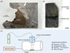 |
Fig. 1 Studied troilite and H+ ion implanter. (a) Seymchan iron meteorite, with a troilite inclusion. (b) Experimental troilite slice with an unirradiated area and an irradiated area. (c) Schematic diagram showing the H+ ion implanter for the solar wind irradiation simulation. |
2 Materials and methods
2.1 Preparation of the sample
The troilite used in the H+ irradiation experiment was sourced from the Seymchan pallasite iron meteorite (Fig. 1a). This meteorite has a low degree of terrestrial weathering and is mainly composed of kamacite, taenite, and troilite (Van Niekerk et al. 2007). The troilite composition was characterized using an electron probe microanalyzer (EPMA); it comprises ~61.8 wt% Fe, ~37.4 wt% S, ~0.71 wt% Co, and ~0.04 wt% Ni. Firstly, the troilite inclusions were separated from this Seymchan meteorite. Then, a slice of troilite of about 4 × 7.5 mm was prepared (Fig. 1b). Finally, it was used to carry out H+ ion irradiation experiments at the Institute of Geochemistry, Chinese Academy of Sciences (Fig. 1c).
2.2 Experimental simulation of solar wind irradiation
H+ ion irradiation was performed on the troilite section using an ion implanter, which consists of a hydrogen gas (H2) source, vacuum system, ionizer, accelerator, vacuum irradiation chamber, and control system (Fig. 1c). This instrument ionized H2 gas and accelerated the ionized H+ ions in an electric field, and then these accelerated H+ ions were implanted into the sample (irradiated area of ~3 cm2) to simulate the solar wind irradiation process. The implanted H+ energy was 1.5 keV, which is commonly used to simulate solar wind H+ irradiation in experiments (Schaible & Baragiola 2014; Tang et al. 2021; Zeng et al. 2021). The average H+ flux was (1.0 ± 0.1) × 1013 ions/cm2/s, similar to fluxes used in other simulations (Loeffler et al. 2008; Christoph et al. 2022; Laczniak et al. 2024), to facilitate the comparison of results (higher flux leads to greater amorphization) (Laczniak et al. 2021, 2024). The total H+ fluence was (1.0 ± 0.1) × 1018 ions/cm2, equivalent to 300 years of irradiation on the lunar surface (Burke et al. 2011) and 1200–3000 years of solar wind H+ implantation in an asteroid belt (2.0–3.3 AU) (Christoph et al. 2022).
Before the experiment, the sample was first dried overnight in the irradiation chamber under vacuum conditions (10−5 Pa). The irradiation was conducted at room temperature (~300 K) and with a pressure of 10−5 Pa for a total duration of 28 hours. To obtain irradiated troilite and unirradiated troilite simultaneously, a ceramic piece was used to partially cover the troilite. At this point, the irradiated and unirradiated areas could be obtained on the same thick troilite section (Fig. 1b).
 |
Fig. 2 Secondary electron images of the unirradiated and irradiated troilite. (a and b) Surface morphology of the unirradiated troilite with different magnifications. (c and d) Surface morphology of the irradiated troilite with different magnifications. |
2.3 Analytical techniques
To quantitatively obtain the chemical composition of the studied troilite, the polished sample, coated with carbon, was measured using the EPMA (JXA-iSP100) at the Macau University of Science and Technology. This sample was measured using the pyrite testing method (Christoph et al. 2022).
The surface morphology and ultrathin foil of the irradiated troilite were analyzed using a Zeiss Crossbeam 350 focused ion beam (FIB) scanning electron microscope (SEM) at the Guangdong University of Technology. For the SEM observations, the conditions were 5 keV voltage and 25–30 000 magnification. Then, a FIB foil was prepared using this instrument. The surface of the working area was coated with two protective Pt layers and then excavated with a Ga ion beam. After obtaining the FIB foil, a low-energy Ga ion beam was used for thinning. The final FIB foil thickness was about 100 nm, and it was welded to the copper grid. This FIB foil was immediately used for transmission electron microscopy (TEM) observation.
The TEM analysis was done using a 200 keV field emission scanning transmission electron microscope (FEI Talos F200S) at the Guangdong University of Technology. Microscopic analysis and crystallographic analysis of the troilite FIB foil were performed using bright field (BF) images, high-angle annular dark field (HAADF) images, and high-resolution transmission electron microscopy (HRTEM), and the Bruker Xflash 6T-30 energy-dispersive X-ray spectroscopy (EDS) detector equipped with the instrument was used to perform chemical nano-analysis.
3 Results
3.1 SEM observations
Scanning electron microscope observations show that there were obvious microscopic modifications on the surface of irradiated troilite compared with the unirradiated troilite (Fig. 2). At a magnification of 10 000 times, it could be vaguely seen that the irradiated area had a rougher texture than the unirradiated area (Figs. 2a and 2c). At a high magnification of 30000 times, it is clearly seen that the surface of irradiated troilite had many dome-like microstructures. These dome-like microstructures are characterized by rounded shapes, nano-scale sizes (~40–100 nm), and a uniform distribution (Fig. 2d).
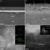 |
Fig. 3 TEM observations of the irradiated troilite. (a) BF-TEM image of the irradiated troilite. (b) BF-TEM image of a close-up view of the irradiated troilite. (c) HRTEM image of unirradiated troilite. (d) HRTEM image of the irradiated layer. |
3.2 TEM analyses
The structure and composition of the H+ irradiated troilite were analyzed using the TEM technique. The BF-TEM image shows that the surface of the troilite was covered with an irradiated layer, with a thickness of ~80–100 nm. In addition, numerous vesicles were observed within the irradiated layer. These vesicles were semicircular and nano-sized (~300 nm in diameter; Fig. 3a). For the crystal structure, the unirradiated troilite had lattice fringes with d-spacing values of 5.2 Å, which correspond to the (0 0 1) crystallographic plane of troilite (Fig. 3c). A fast Fourier transform (FFT; top-right corners of Figs. 3c and 3d) of the HRTEM image shows that the irradiated troilite changed from a single-crystalline to a polycrystalline state (Fig. 3d). In the unirradiated region (Fig. 3c), the FFT of the single-crystalline troilite showed discrete diffraction spots arranged regularly, and the distribution of these spots corresponds to the Bragg diffraction conditions of its crystal structure. In the irradiated region (Fig. 3d), the FFT of the polycrystalline troilite showed a coexistence of diffraction spots and diffraction rings. This is due to the presence of small single-crystalline grains with different orientations in the selected area, resulting in a polycrystalline structure.
The d-spacings of the low-index diffraction spots of unirradiated single-crystalline troilite were measured as A = 5.2 Å, B = 3.0 Å, C = 5.2 Å, and D = 5.2 Å, with the inter-planar angles between these low-index diffraction spots shown in the figure. In the FFT image of the polycrystalline region (Fig. 3d), distinct polycrystalline diffraction rings corresponding to the (1 1 0) plane (d-spacing = 3.0 Å) and the (0 3 0) plane (d-spacing = 1.7 Å) can be clearly observed. While typical polycrystalline diffraction signatures should display continuous concentric rings, the FFT image of the polycrystalline region in Figure 3d shows a partially closed but incomplete ring pattern, indicating that the irradiated area has not yet fully transitioned to a polycrystalline state and that the analyzed area contains a small number of grains.
In addition, TEM-EDS analysis reveals that there were obvious composition changes in the irradiated layer of troilite (Figs. 4 and 5). Compared to the unirradiated troilite, the irradiated layer of troilite contained less S content (Fig. 4b). Specifically, the intensities of S and Fe begin to weaken at a depth of 100 nm in the irradiated layer (Fig. 4b). The S content gradually decreases from the inside of the irradiated layer to the outermost of the irradiated layer (i.e., ranging from ~37 wt% to ~5 wt%; Fig. 4c). The vesicle-bearing irradiated layer shows a similar chemical trend as the vesicle-free irradiated layer (Fig. 5). No chemical changes were observed in the vesicles.
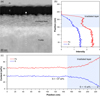 |
Fig. 4 Chemical variation in the irradiated layer of troilite. (a) HAADF image of an irradiated layer of troilite. The dashed arrow indicates the position of the TEM-EDS line scan. (b) TEM-EDS line scan results. (c) Fe and S content (wt%) changes in the irradiated layer of troilite. |
4 Discussion
4.1 Comparison with the space-weathered troilite in return samples
The space-weathered troilite in return samples are commonly characterized by the presence of iron whisker, sulfur loss in the irradiated layer, and vesicles (Matsumoto et al. 2020, 2021; Chaves & Thompson 2022; Li et al. 2024a). For the H+ irradiation experiments, no iron whisker was found, unlike that in the lunar and Itokawa asteroid samples (Matsumoto et al. 2020, 2021). However, an obvious sulfur loss in the irradiated layer of troilite was observed (Fig. 4). The irradiated layer (Fig. 3b, ~80 nm) in troilite caused by experimental H+ irradiation was significantly thicker than the rim of troilite (~10 nm) observed in asteroid return samples (Harries & Langenhorst 2014; Matsumoto et al. 2020; Chaves & Thompson 2022). In the absence of significant heating or a possible vapor-deposited layer source, the cause of troilite polycrystallization is likely related to significant sulfur mass loss, as observed in the returned samples from Itokawa (Harries & Langenhorst 2014). Additionally, some vesicles were also observed in the H+ irradiated layer (Figs. 3a and 5a), like those in the lunar and Itokawa asteroid samples (Matsumoto et al. 2020, 2021). The S content of these vesicles remained unchanged (Fig. 5c), which is consistent with that in the Itokawa regolith particles (Matsumoto et al. 2020).
For the troilite grains in Apollo soil, the depth (~80–100 nm) of their irradiated layer (Matsumoto et al. 2021) is similar to the result of our 300-year solar wind irradiation simulation (i.e., ~80 nm; Figs. 3b, 4a, and 5a). The irradiated layer is deeper than the ion penetration depths calculated by the Monte Carlo-based Stopping and Range of Ions in Matter (SRIM) simulation for FeS (Matsumoto et al. 2020, 2021) (Fig. B.1). The mineral damage effects that SRIM cannot simulate, such as the differences in mineral damage caused by different ion irradiation fluxes (Laczniak et al. 2024) and radiation-enhanced diffusion due to particle interactions in target materials (Doyle et al. 2018), may lead to an underestimation of the mineral damage caused by the solar wind on troilite. The vesicles formed beneath the irradiation layer (Figs. 3a and 5a) also suggest the formation of deep vesicles in returned samples, and their internal compositions might be associated with H+ irradiation (Matsumoto et al. 2021). Additionally, similar to the partial polycrystallization observed in the simulation experiments, the space-weathered troilite in the Apollo return samples is crystallographically altered. The crystal plane arrangement of the troilite space-weathered rim shows a distortion (nonlinear alignment) in the HRTEM images (Matsumoto et al. 2021).
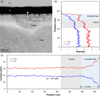 |
Fig. 5 Chemical variation in the irradiated layer (with vesicles) of troilite. (a) HAADF image of an irradiated layer (with vesicles) of troilite. The dashed arrow indicates the position of the TEM-EDS line scan. (b) TEM-EDS line scan results. (c) Fe and S content (wt%) changes in the irradiated layer (with vesicles) of troilite. |
4.2 Loss of sulfur on troilite induced by space weathering
Our experimental results show that the loss of sulfur on irradiated troilite is not linear (i.e., the sulfur loss is more severe closer to the exposure surface; Figs. 4c and 5c). During the simulated 300-year irradiation period of the lunar surface, we estimated that the mass loss of sulfur on the irradiated layer of troilite is ~32 wt% (equivalent to ~0.1 wt%/yr). For the vesicles within the irradiated layer of troilite, no significant sulfur loss was observed in this region (Fig. 5c). This is consistent with observations of the Itokawa asteroid regolith (Matsumoto et al. 2020). Specifically, the mass loss of sulfur in the irradiated layer above vesicles is about 32 wt%, which is consistent with the vesicle-free regions (Fig. 4c). This implies that the vesicles do not affect the sulfur loss rate in the irradiation layer of troilite. We therefore suggest that these vesicles formed due to structural damage from H+ ion implantation.
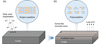 |
Fig. 6 Schematic diagram showing the space-weathering effect on troilite from solar wind H+ irradiation. The morphological, crystal, and chemical changes before H+ irradiation (a) and after H+ irradiation (b) are shown. |
4.3 Implications for space weathering on airless planetary bodies
Space-weathered silicate minerals are commonly covered by an amorphous layer damaged by solar wind H+ ion implantation or the micrometeorite bombardment thermal effect, which are also believed to be related to the growth mechanisms of troilite-iron whiskers. The growth of troilite-iron whiskers induced by thermal alteration has been confirmed through molecular dynamics simulations (Li et al. 2024b) and in situ simulated heating experiments (Thompson et al. 2025), providing robust evidence of the involvement of thermal effects in troilite space-weathering alteration. Although current simulation experiments based on H+ ion irradiation have not successfully replicated iron whisker structures, this may be due to the limited timescale (hundreds to thousands of years) simulated for space weathering via ion implantation (Loeffler et al. 2008; Christoph et al. 2022).
Under H+ ion implantation, sputtering of S and Fe in troilite occurs, generating lattice defects (Matsumoto et al. 2020, 2021; Christoph et al. 2022). The diffusion of these defects within the polycrystalline layer facilitates the formation of higher-order defects, thereby accelerating polycrystallization (Christoph et al. 2022). The preferential sputtering of S leads to significant sulfur loss in the irradiated layer, coupled with cation super-saturation that drives cation diffusion (Matsumoto et al. 2020; Christoph et al. 2022; Thompson et al. 2025). Additionally, H+ ions react with S to form H2S, which escapes in gaseous form, further contributing to sulfur depletion (Harries & Langenhorst 2014; Matsumoto et al. 2020, 2021). This study confirms that the effects of solar wind irradiation on troilite can cause both microstructural and chemical alterations, which are summarized in the schematic diagram of Figure 6.
From a simulation perspective, thermal effects and ion implantation cause different modification characteristics on troilite. Thus, space-weathering features in troilite can be used to distinguish between these driving mechanisms. Transient high-temperature thermal effects result in space-weathered troilite with clean single-crystalline boundaries between troilite and weathering products (e.g., iron whiskers), which is a sign of recrystallization (Li et al. 2024b; Thompson et al. 2025). In contrast, prolonged heating triggers outward S diffusion, leading to surface sulfur enrichment (Thompson et al. 2025). The space-weathered troilite without iron whisker formation found in the Itokawa return sample shows a different response, one that includes polycrystallization and sulfur loss from the troilite surface (Harries & Langenhorst 2014; Chaves & Thompson 2022); this is likely related to solar wind irradiation, as it is consistent with the results of our ion implantation simulation experiments. Therefore, this work provides experimental evidence of varying space-weathering textures in troilite induced by micrometeorite impacts versus changes due to solar wind irradiation.
Troilite is a commonly distributed accessory mineral on the surface of the Moon and asteroids (Evans Jr 1970; Kemper et al. 2004; Skala et al. 2006; Li et al. 2023). Our work shows that the sulfur mass loss rate of troilite under solar wind irradiation is ~0.1 wt%/yr. This means that large amounts of sulfur can continue to be lost from the surface of airless planetary bodies through widespread space-weathering effects. We have estimated the sulfur loss from troilite by solar wind H+ irradiation on the Moon to be 4.5 × 10¹⁰ kg/yr (Appendix A). If correct, this indicates that solar wind H+ ion implantation is a driving factor affecting the composition, distribution, and circulation of sulfur on the Moon and other airless planetary bodies in the Solar System.
5 Conclusions
In this work we investigated the chemical and mineralogical changes in troilite induced by space weathering through simulated solar wind H+ irradiation. This study provides the first quantitative analysis of such microstructural alterations in troilite under simulated space-weathering conditions. Our results reveal that solar wind irradiation significantly modifies the morphological, chemical, and structural properties of troilite. Dome-like microstructures formed on the irradiated troilite surface, and sulfur depletion was observed in the irradiated layer. Based on our measurements, the estimated mass loss rate of sulfur due to H+ irradiation is 0.1 wt%/yr. Crystallographically, the irradiated troilite transformed from a single-crystalline to a polycrystalline state. Future studies should explore additional space-weathering effects, such as micrometeorite bombardment, as potential drivers of troilite microstructural alteration on airless planetary bodies. Furthermore, the influence of space weathering on sulfur isotope fractionation in troilite warrants further investigation.
Acknowledgements
This work is supported by National Natural Science Foundation of China (No. 42241104), Science and Technology Development Fund (FDCT) of Macau (grant Nos. 0014/2022/A1, 0034/2024/AMJ, 0008/2024/AKP, 002/2024/SKL). The authors are grateful to the reviewer Dr. John Christoph for his recommendations and comments, which improved this paper significantly.
Appendix A Estimation of sulfur loss by solar wind H+ irradiation on the Moon
The sulfur loss on the Moon is estimated from the following equation:
 (A.1)
(A.1)
where:
Sloss is the sulfur loss driven by solar wind H+ irradiation
Ctr is the troilite content in lunar basaltic regolith
Rs represents the sulfur mass loss rate of troilite under solar wind H+ irradiation
These parameters are estimated as follows:
Ctr = Total mass of lunar basaltic regolith
(4.5 × 10¹⁶ kg; (Heiken et al. 1991))
× Troilite abundance in regolith
(0.1 wt%; (Steenstra & van Westrenen 2017; Wang et al. 2024))
-
= 4.5 × 10¹³ kg

Therefore, the sulfur loss by solar wind H+ irradiation on the Moon is approximately 4.5 × 10¹⁰ kg/yr.
Appendix B SRIM simulation of solar wind ion implantation depth distribution in troilite
The implantation depth of solar wind ions was simulated using the SRIM software. The simulation employed 100000 particles implanted into troilite (FeS, density 4.69 g/cm3) under the “Monolayer Collision Steps / Surface Sputtering” mode. The Y-axis represents ion concentration, and the X-axis corresponds to the surface depth of troilite. The ion penetration depth, defined as the sum of the average projected range and the longitudinal straggle (Matsumoto et al. 2020), is approximately 22 nm, 30 nm, and 47 nm for 1 keV H, 1.5 keV H, and 4 keV He, respectively.
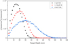 |
Fig. B.1 SRIM simulations of the depth distribution for implantation of different-energy particles in troilite. |
References
- Anand, M., Taylor, L. A., Nazarov, M. A., et al. 2004, PNAS, 101, 6847 [Google Scholar]
- Bennett, C. J., Pirim, C., & Orlando, T. M. 2013, Chem. Rev., 113, 9086 [NASA ADS] [CrossRef] [Google Scholar]
- Binzel, R. P., Morbidelli, A., Merouane, S., et al. 2010, Nature, 463, 331 [NASA ADS] [CrossRef] [Google Scholar]
- Burgess, K., & Stroud, R. 2018, J. Geophys. Res.: Planets, 123, 2022 [Google Scholar]
- Burke, D., Dukes, C., Kim, J.-H., et al. 2011, Icarus, 211, 1082 [Google Scholar]
- Chaves, L. C., & Thompson, M. S. 2022, Earth Planets Space, 74, 124 [Google Scholar]
- Christoffersen, R., & Keller, L. P. 2011, Meteor. Planet. Sci., 46, 950 [Google Scholar]
- Christoph, J., Minesinger, G., Bu, C., Dukes, C., & Elkins-Tanton, L. 2022, J. Geophys. Res.: Planets, 127, e2021JE006916 [Google Scholar]
- Doyle, P. J., Benensky, K. M., & Zinkle, S. J. 2018, J. Nucl. Mater., 509, 168 [Google Scholar]
- Dukes, C., Baragiola, R., & McFadden, L. 1999, J. Geophys. Res.: Planets, 104, 1865 [Google Scholar]
- Evans Jr, H. T. 1970, Science, 167, 621 [Google Scholar]
- Fulvio, D., Maron, L. F., Perez, Y. C., et al. 2021, Icarus, 366, 114532 [CrossRef] [Google Scholar]
- Greer, J., Rout, S. S., Isheim, D., et al. 2020, Meteor. Planet. Sci., 55, 426 [Google Scholar]
- Gu, L., Chen, Y., Xu, Y., et al. 2022, Geophys. Res. Lett., 49, e2022GL097875 [Google Scholar]
- Guo, Z., Li, C., Li, Y., et al. 2022a, Geophys. Res. Lett., 49, e2021GL097323 [Google Scholar]
- Guo, Z., Li, C., Li, Y., et al. 2022b, Nat. Commun., 13, 7177 [Google Scholar]
- Hapke, B. 2001, J. Geophys. Res.: Planets, 106, 10039 [NASA ADS] [CrossRef] [Google Scholar]
- Harries, D., & Langenhorst, F. 2014, Earth Planets Space, 66, 1 [CrossRef] [Google Scholar]
- Heiken, G., Vaniman, D., & French, B. M. 1991, Lunar Sourcebook: A User’s Guide to the Moon, No. 1259 (Cup Archive) [Google Scholar]
- Keller, L. P., & Berger, E. L. 2014, Earth Planets Space, 66, 1 [CrossRef] [Google Scholar]
- Keller, L. P., & McKay, D. S. 1993, Science, 261, 1305 [NASA ADS] [CrossRef] [Google Scholar]
- Keller, L. P., & McKay, D. S. 1997, Geochim. Cosmochim. Acta, 61, 2331 [NASA ADS] [CrossRef] [Google Scholar]
- Keller, L., Rahman, Z., Hiroi, T., et al. 2013, in Lunar and Planetary Science Conference, No. JSC-CN-28006 [Google Scholar]
- Kemper, F., Vriend, W., & Tielens, A. 2004, ApJ, 609, 826 [Google Scholar]
- Laczniak, D., Thompson, M., Christoffersen, R., et al. 2021, Icarus, 364, 114479 [CrossRef] [Google Scholar]
- Laczniak, D., Thompson, M., Christoffersen, R., et al. 2024, Icarus, 410, 115883 [Google Scholar]
- Li, Y., Li, X., Wang, S., et al. 2013, J. Geophys. Res.: Planets, 118, 1974 [NASA ADS] [CrossRef] [Google Scholar]
- Li, X., Chen, Y., Tang, X., et al. 2023, Icarus, 390, 115299 [Google Scholar]
- Li, C., Li, Y., Wei, K., et al. 2024a, Geochim. Cosmochim. Acta, 387, 28 [Google Scholar]
- Li, C., Li, Y., Wei, K., et al. 2024b, Geochim. Cosmochim. Acta, 379, 134 [Google Scholar]
- Loeffler, M., Dukes, C., Chang, W., McFadden, L., & Baragiola, R. 2008, Icarus, 195, 622 [Google Scholar]
- Loeffler, M., Dukes, C., Christoffersen, R., & Baragiola, R. 2016, Meteor. Planet. Sci., 51, 261 [NASA ADS] [CrossRef] [Google Scholar]
- Marchi, S., Black, B. A., Elkins-Tanton, L. T., & Bottke, W. F. 2016, Earth Planet. Sci. Lett., 449, 96 [Google Scholar]
- Matsumoto, T., Harries, D., Langenhorst, F., Miyake, A., & Noguchi, T. 2020, Nat. Commun., 11, 1117 [Google Scholar]
- Matsumoto, T., Noguchi, T., Tobimatsu, Y., et al. 2021, Geochim. Cosmochim. Acta, 299, 69 [Google Scholar]
- McCoy, T. J., Burbine, T., McFadden, L., et al. 2001, Meteor. Planet. Sci., 36, 1661 [Google Scholar]
- Nittler, L. R., Starr, R. D., Lim, L., et al. 2001, Meteor. Planet. Sci., 36, 1673 [Google Scholar]
- Noguchi, T., Nakamura, T., Kimura, M., et al. 2011, Science, 333, 1121 [NASA ADS] [CrossRef] [Google Scholar]
- Pang, R., Li, Y., Li, C., et al. 2024, Acta Geochim., 43, 774 [Google Scholar]
- Pieters, C. M., & Noble, S. K. 2016, J. Geophys. Res.: Planets, 121, 1865 [NASA ADS] [CrossRef] [Google Scholar]
- Prince, B., Magnuson, M., Chaves, L., Thompson, M., & Loeffler, M. 2020, J. Geophys. Res.: Planets, 125, e2019JE006242 [Google Scholar]
- Schaible, M. J., & Baragiola, R. A. 2014, J. Geophys. Res.: Planets, 119, 2017 [NASA ADS] [CrossRef] [Google Scholar]
- Skala, R., Cisarova, I., & Drabek, M. 2006, Am. Mineralogist, 91, 917 [Google Scholar]
- Steenstra, E. S., & van Westrenen, W. 2017, in Encyclopedia of Lunar Science (Springer), 1 [Google Scholar]
- Tang, H., Li, X., Zeng, X., et al. 2021, Icarus, 359, 114322 [NASA ADS] [CrossRef] [Google Scholar]
- Thompson, M. S., Zega, T. J., Becerra, P., Keane, J. T., & Byrne, S. 2016, Meteor. Planet. Sci., 51, 1082 [Google Scholar]
- Thompson, M. S., Davidson, J., Schrader, D. L., & Zega, T. J. 2025, Nat. Commun., 16, 5975 [Google Scholar]
- Trombka, J., Squyres, S. W., Bruckner, J., et al. 2000, Science, 289, 2101 [Google Scholar]
- Van Niekerk, D., Greenwood, R., Franchi, I., Scott, E., & Keil, K. 2007, Meteor. Planet. Sci. Suppl., 42, 5196 [Google Scholar]
- Wang, Z., Li, Y., Zhang, W., et al. 2024, Geochim. Cosmochim. Acta, 368, 168 [Google Scholar]
- Xu, J., Mo, B., Wu, Y., et al. 2023, A&A, 672, A115 [NASA ADS] [CrossRef] [EDP Sciences] [Google Scholar]
- Zeng, X., Tang, H., Li, X., et al. 2021, Earth Planet. Sci. Lett., 560, 116806 [CrossRef] [Google Scholar]
- Zeng, X., Wu, Y., Yu, W., et al. 2024, Nat. Astron., 8, 732 [Google Scholar]
All Figures
|
Fig. 1 Studied troilite and H+ ion implanter. (a) Seymchan iron meteorite, with a troilite inclusion. (b) Experimental troilite slice with an unirradiated area and an irradiated area. (c) Schematic diagram showing the H+ ion implanter for the solar wind irradiation simulation. |
| In the text | |
|
Fig. 2 Secondary electron images of the unirradiated and irradiated troilite. (a and b) Surface morphology of the unirradiated troilite with different magnifications. (c and d) Surface morphology of the irradiated troilite with different magnifications. |
| In the text | |
|
Fig. 3 TEM observations of the irradiated troilite. (a) BF-TEM image of the irradiated troilite. (b) BF-TEM image of a close-up view of the irradiated troilite. (c) HRTEM image of unirradiated troilite. (d) HRTEM image of the irradiated layer. |
| In the text | |
|
Fig. 4 Chemical variation in the irradiated layer of troilite. (a) HAADF image of an irradiated layer of troilite. The dashed arrow indicates the position of the TEM-EDS line scan. (b) TEM-EDS line scan results. (c) Fe and S content (wt%) changes in the irradiated layer of troilite. |
| In the text | |
|
Fig. 5 Chemical variation in the irradiated layer (with vesicles) of troilite. (a) HAADF image of an irradiated layer (with vesicles) of troilite. The dashed arrow indicates the position of the TEM-EDS line scan. (b) TEM-EDS line scan results. (c) Fe and S content (wt%) changes in the irradiated layer (with vesicles) of troilite. |
| In the text | |
|
Fig. 6 Schematic diagram showing the space-weathering effect on troilite from solar wind H+ irradiation. The morphological, crystal, and chemical changes before H+ irradiation (a) and after H+ irradiation (b) are shown. |
| In the text | |
|
Fig. B.1 SRIM simulations of the depth distribution for implantation of different-energy particles in troilite. |
| In the text | |
Current usage metrics show cumulative count of Article Views (full-text article views including HTML views, PDF and ePub downloads, according to the available data) and Abstracts Views on Vision4Press platform.
Data correspond to usage on the plateform after 2015. The current usage metrics is available 48-96 hours after online publication and is updated daily on week days.
Initial download of the metrics may take a while.